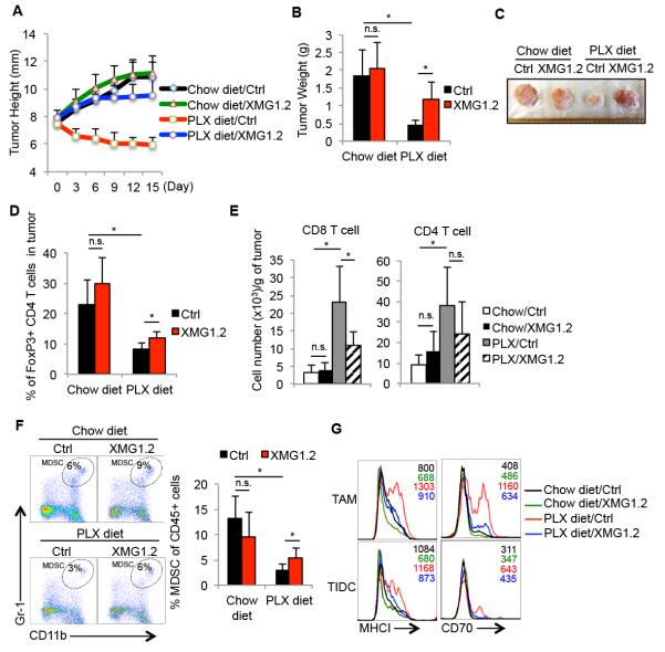
Figure 6. PLX4720 inhibits intratumoral accumulation of myeloid suppressor cells in melanomas in an IFNγ-dependent mechanism.
Five weeks after tumor induction, BrafV600E/Pten melanoma-bearing mice were fed normal or PLX-containing chow for another 15 days. Control vehicle or IFNγ-neutralizing mAb (XMG1.2) was injected i.p. every three days during this 15-day period and the growth rate (A), weight (B), and appearance (C) of the tumors were assessed. (D) FACS plots (left) and bar graphs (right) show the percentage of FoxP3+ CD4+ Tregs in tumors and spleens post treatment as measured by flow cytometry. Data shown are pooled from 2 independent experiments that contain in total 7-12 mice per group. (E-F) Bar graphs show the cell number of tumor infiltrating T cells (n=5-8 mice per group) (E) or percentage of MDSCs (F) post treatment as measured by flow cytometry (n=7-11 mice per group). (G) Histograms show the expression of indicated maturation markers on the TAMs and TIDCs isolated from the tumors following treatment. *, P<0.05 in unpaired student t test. n.s.= not significant in unpaired student t test.

