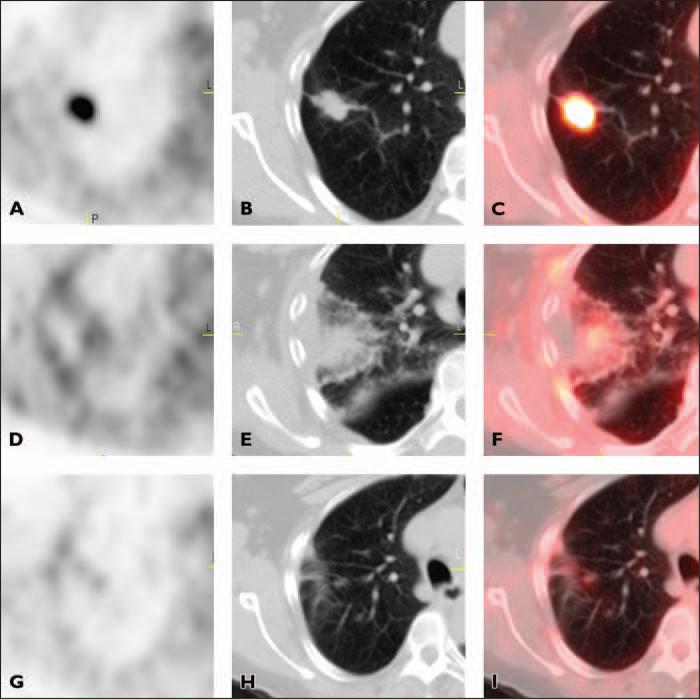
Fig. 2.
66-year-old woman with stage IA non–small cell lung cancer. A–C, Axial PET (A), CT (B), and fused PET/CT (C) images obtained before radiofrequency ablation (RFA) show area of intense FDG uptake corresponding to tumor in right upper lobe.
D–F, Axial PET (D), CT (E), and fused PET/CT (F) images obtained 3 days after RFA show homogeneous rim activity along periphery of ablation site with central photopenia (complete response). Rim activity is higher than background mediastinal activity.
G–I, Axial PET (G), CT (H), and fused PET/CT (I) images 6 months after RFA no longer show homogeneous rim activity. Mild linear FDG uptake at ablation site has intensity similar to mediastinal blood pool activity (complete response).

