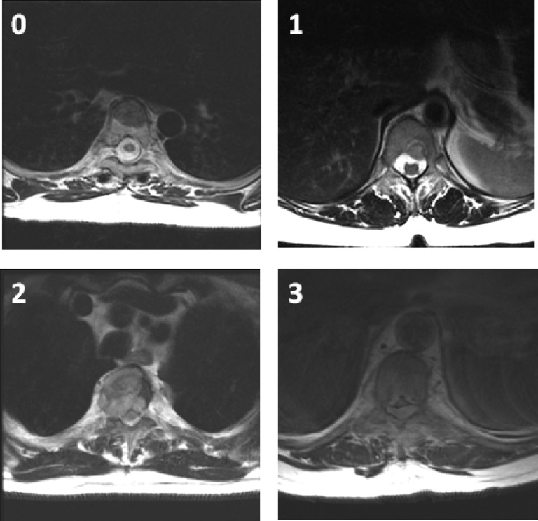
Figure 1.
A six-point grading system by the Spine Oncology Study Group [5] uses axial T2-weighted images at the site of most severe compression to describe the degree of epidural spinal cord compression: 0, tumor is confined to bone only; 1, tumor extension into the epidural space without deformation of the spinal cord; 2, spinal cord compression but cerebrospinal fluid is visible; and 3, spinal cord compression without visible cerebrospinal fluid. The grade 1 delineation is further subdivided into 1a–1c: 1a, epidural impingement but no deformation of the thecal sac; 1b, deformation of the thecal sac but without spinal cord abutment; and 1c, deformation of the thecal sac with spinal cord abutment but without compression.

