Figure 4.
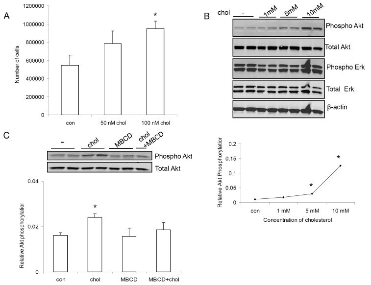
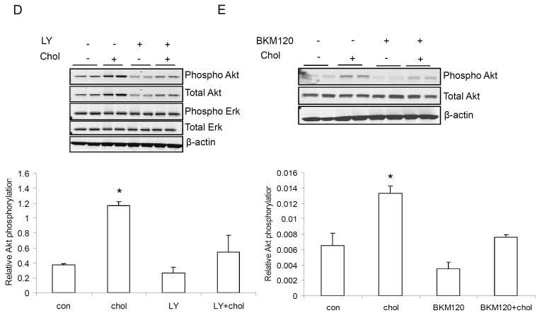
100 nM (equivalent to 4 μg/dl) cholesterol significantly increased the number of Mvt-1 cells in vitro (A). Cholesterol stimulated AktS473 phosphorylation in Mvt-1 cells in a dose dependent manner (1-10 mM equivalent to 40-400 mg/dl) (B). 1mM of cholesterol activated Akt during a shorter time period (20 min and 40 min) (C). MBCD is known to deplete cholesterol from plasma membrane leading to disruption of lipid rafts. The presence of MBCD (5 mM) inhibited cholesterol-induced AktS473 phosphorylation (D). Densitometric analysis of Akt phosphorylation is presented as levels of phosphorylation compared to total Akt protein. Statistically significant differenced is indicated as *, P<0.05, Student’s t test. (*, P<0.05 untreated vs cholesterol treated Mvt-1 cells and/or cholesterol treated vs cholesterol and inhibitor treated Mvt-1 cells). Statistical analyses were determined using the student’s t test where 2 groups were present and ANOVA for comparing more than 2 groups.


