Figure 3.

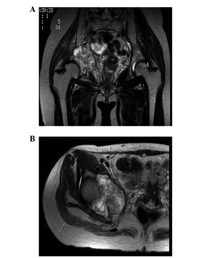
Case one: (A) Coronal and (B) axial T2-weighted magnetic resonance imaging (MRI) showing a mass with inhomogeneous intensity located in the right acetabulum.

Case one: (A) Coronal and (B) axial T2-weighted magnetic resonance imaging (MRI) showing a mass with inhomogeneous intensity located in the right acetabulum.