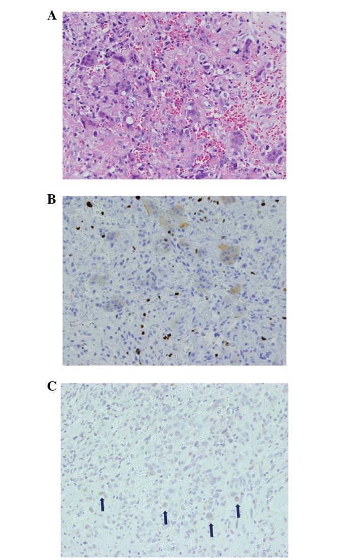
Figure 4.

Case one: Open biopsy of the acetabular lesion. (A) Histological examination revealed spindle and round cells, with the multinucleated giant cells in a collagenous matrix with capillaries (hematoxylin-eosin stain; magnification, ×400). In the immunological studies (B) the Ki-67 index was 20% and (C) the tumor cells were fibroblast growth factor 23-positive (indicated by the arrows; immunohistochemical stain; magnification, ×200). Based on these observations, the tumor was diagnosed as a malignant phosphaturic mesenchymal tumor (PMT).
