Figure 2.
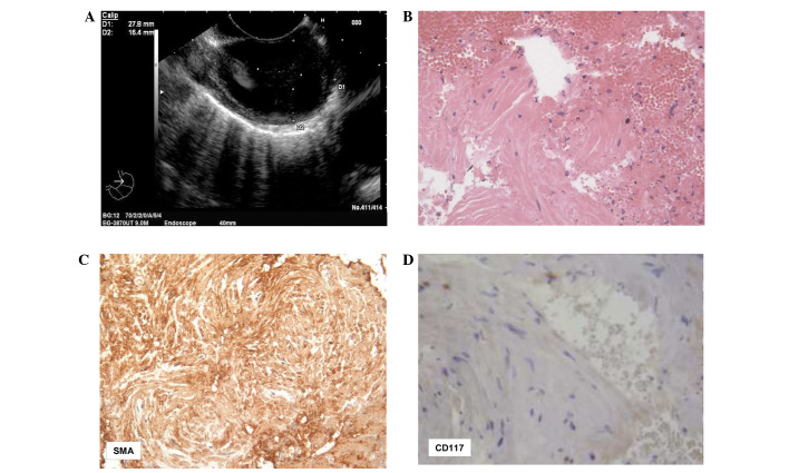
Case 2: (A) EUS scanning results revealing a 27.8×16.4-mm, hypoechoic, ovoid, well-delimited lesion originating from the muscle layer of the distal esophagus. (B) Clusters of spindle cells intermingled with red blood cells are indicative of leiomyoma (hematoxylin and eosin staining; magnification, ×160). (C) These elements were reactive for SMA (immunoperoxidase and Mayer’s hemalum counterstain; magnification, ×120), (D) while no immunoreactivity was found with CD117 (immunoperoxidase and Mayer’s hemalum counterstain; magnification, ×160). EUS, endoscopic ultrasound; SMA, smooth muscle actin.

