
Figure 1.
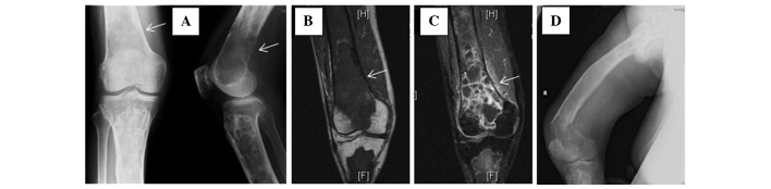
(A) Roentgenograms showing typical fibrous dysplasia involving the tibia and an ill-defined osteolytic lesion (arrow) in the right distal femur. (B) T1-weighted coronal-plane magnetic resonance imaging of the lesion of secondary osteosarcoma (arrow). (C) The lesion was intensively enhanced by injection of gadopentetate dimeglumine (arrow). (D) A pathological fracture through the lesion of the distal femur.