Figure 2.

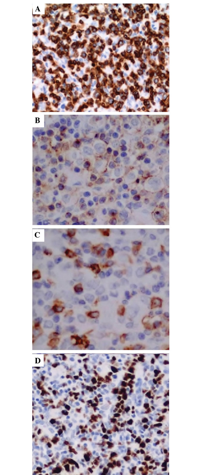
Immunohistochemical analysis of the lymph node in the inguinal fold: (A) Cluster of differentiation (CD)3-positive; (B) CD2-positive; (C) CD5-positive; and (D) Ki-67-positive (hematoxylin-eosin stain; magnification, ×200).

Immunohistochemical analysis of the lymph node in the inguinal fold: (A) Cluster of differentiation (CD)3-positive; (B) CD2-positive; (C) CD5-positive; and (D) Ki-67-positive (hematoxylin-eosin stain; magnification, ×200).