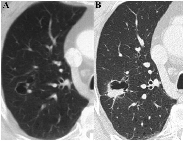
Figure 6.
Male, 61 years old. The patient presented with (A) a cystic cavity in the posterior segment of the right lung upper lobe, no compartment, a slightly thickened wall and punctiform wall nodules. (B) Re-examination at month 18 demonstrated that the cavity was marginally enlarged, the wall was thickened, and the wall nodules were clearly enlarged and moderately enhanced. In addition, no compartments in the cavity were observed.

