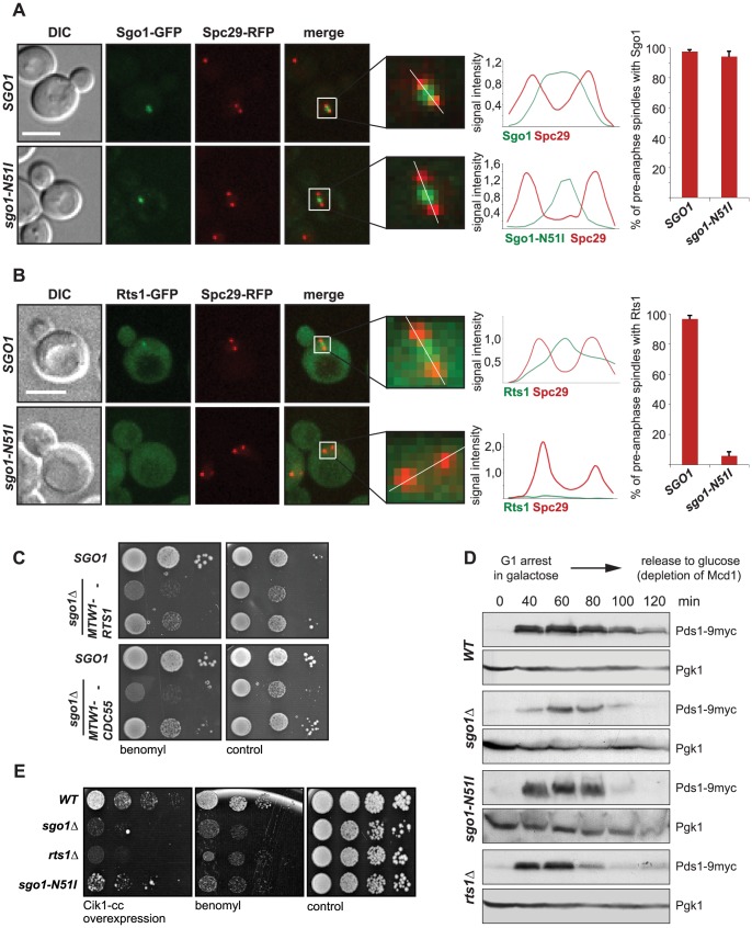
Figure 1. Sgo1-mediated PP2A recruitment to the centromere is essential for tension sensing.
(A) Localization of the GFP-tagged Sgo1 and Sgo1-N51I mutant. Spindle pole bodies (SPBs) are visualized with Spc29-RFP. Only pre-anaphase spindles (SPB distance <2 µm, spindle located in the mother cell) were scored. Plots on the right: histogram of signal intensity across the white line in the insets and percentage of cells with localized GFP signal. Bar – 5 µm. (B) Localization of Rts1-GFP in wild type cells or in cells carrying the sgo1-N51I mutation. SPBs are visualized with Spc29-RFP. Only pre-anaphase spindles (SPB distance <2 µm, spindle located in the mother cell) were scored. Plots on the right: histogram of signal intensity across the white line in the insets and percentage of cells with localized GFP signal. Bar – 5 µm. (C) Sensitivity to the microtubule depolymerizing drug benomyl in cells lacking SGO1. Cell viability was scored upon artificial tethering of the PP2A regulatory subunit Rts1 and Cdc55, respectively, to the kinetochore. (D) Progression of the wild type, sgo1Δ, sgo1-N51I and rts1Δ mutants through cell cycle upon depletion of the cohesin subunit Mcd1 which leads to formation of tensionless kinetochores. (E) Sensitivity of the wild type, sgo1Δ, rts1Δ and sgo1-N51I mutants to microtubule poisons and to the overexpression of Cik1-cc which triggers the formation of syntelic attachments at high frequencies.

