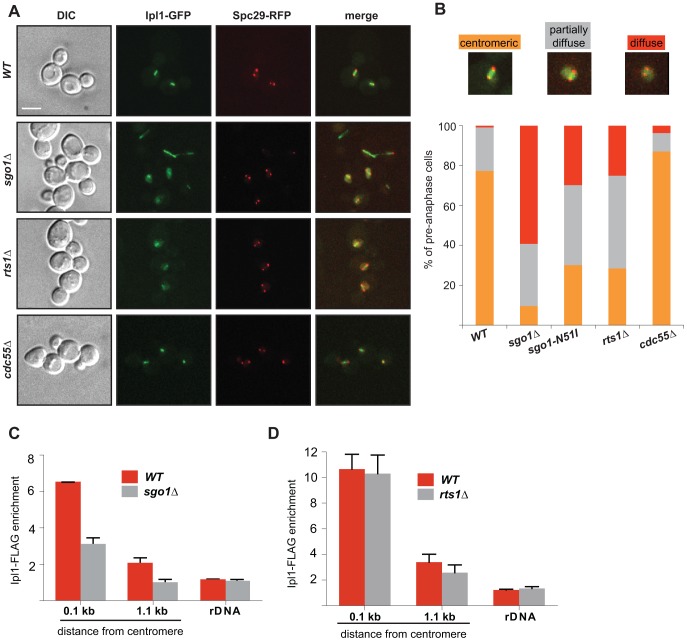
Figure 4. Sgo1 and Rts1 are required for the maintenance of Ipl1 localization and activity at the centromere.
(A) Localization of Ipl1-GFP during mitosis in wild type cells and in cells lacking Sgo1, Rts1 or Cdc55. SPBs are visualized with Spc29-RFP. Bar – 5 µm. (B) Quantification of Ipl1-GFP localization on pre-anaphase spindles in wild type and sgo1Δ, sgo1-N51I, rts1Δ and cdc55Δ mutants (SPB distance <2 µm). Mean values of three independent experiments are shown. At least 150 cells were scored in each experiment. Top – examples of scored categories. (C) Enrichment of Ipl1-FLAG on centromeric DNA (0.1 kb away from CEN1 and 1.1 kb away from CEN4) and on rDNA (NTS1-2) normalized to the levels of Ipl1-FLAG bound to the arm of chromosome 10 in mitotic cells. ChIP-qPCR experiments of Ipl1-FLAG were performed using wild type and sgo1Δ cells arrested with nocodazole. Error bars represent the standard error of the mean. (D) Enrichment of Ipl1-FLAG on centromeric DNA (0.1 kb away from CEN1 and 1.1 kb away from CEN4) and on rDNA (NTS1-2) normalized to the levels of Ipl1-FLAG bound to the arm of chromosome 10 in mitotic cells. ChIP-qPCR experiments of Ipl1-FLAG were performed using wild type and rts1Δ cells arrested with nocodazole. Error bars represent the standard error of the mean.

