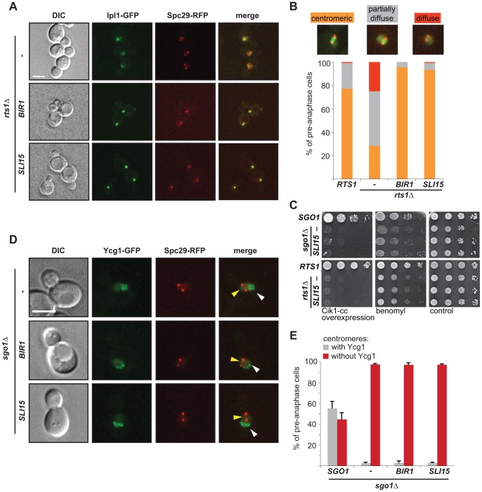
Figure 5. Dual role of Sgo1 in localization of condensin and Ipl1.
(A) Localization of Ipl1-GFP in rts1Δ cells that overexpress BIR1 or SLI15. SPBs are visualized with Spc29-RFP. Bar – 5 µm. (B) Quantification of Ipl1-GFP localization on pre-anaphase spindles. Means of three independent experiments are shown. At least 150 cells were scored in each experiment. (C) Growth of sgo1Δ and rts1Δ cells which overexpress BIR1 or SLI15 in the presence of microtubule poisons and under conditions leading to syntelic attachments. (D) Localization of the Ycg1-GFP signal in sgo1Δ cells which overexpress BIR1 or SLI15. SPBs are visualized with Spc29-RFP. Yellow arrowheads indicate lack of centromeric localization. White arrowheads indicate Ycg1-GFP localized to rDNA. Bar – 5 µm. (E) Quantification of Ycg1-GFP localization. Only pre-anaphase spindles were scored (SPB distance <2 µm). Means with SD of three independent experiments are shown. At least 150 cells were scored in each experiment.

