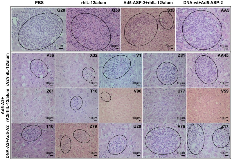
Figure 7. Histopathological analysis of liver from macaques at 6 weeks post-infection with L. infantum.
Photomicrographs of liver from representative macaques from control (PBS; rhIL-12/alum; Ad5-ASP-2+rhIL-12/alum; and DNA-wt+Ad5-ASP-2) as well as vaccinated (rA2/rhIL-12/alum; Ad5-A2+rA2/rhIL-12/alum; DNA-A2+Ad5-A2) macaques at 6 weeks post-infection. In the top panel (control groups), sections showing multifocal coalescing hepatic immune granulomas (circled), consisting of an aggregation of activated macrophages, surrounded by lymphocytes, which obliterate the sinusoids and protrude the parenchyma. Also illustrated are proliferation and hyperplasia of parasite-laden Kupfer cells (arrows), associated with fatty changes in stellate cells (arrow-heads). In the other panels (vaccinated animals of groups rA2/rhIL-12/alum, Ad5-A2+rA2/rhIL-12/alum and DNA-A2+Ad5-A2), sections show functional (parasite-free) hepatic granulomas (circled), which were much reduced in number and size (compared to those of the controls) at this stage of infection.

