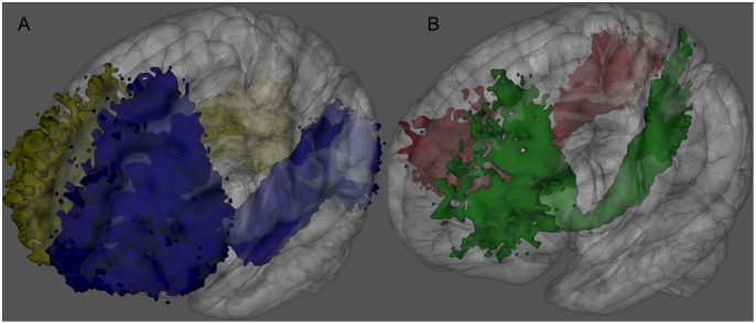
Figure 2. IFOFq subdivision into portions connecting frontal regions to either the occipital (A) or the parietal lobes (B).
3D rendering of the left (blue) and right (yellow) IFOFocc reconstructed on the 20 subjects normalized to the MNI space (A). 3D rendering of the left (green) and right (red) portions of the IFOFq connecting frontal and parietal lobes reconstructed on the 20 subjects normalized to the MNI space (B). Voxels that were visited in the residual bootstrap tractography in at least 20% of subject are shown.

