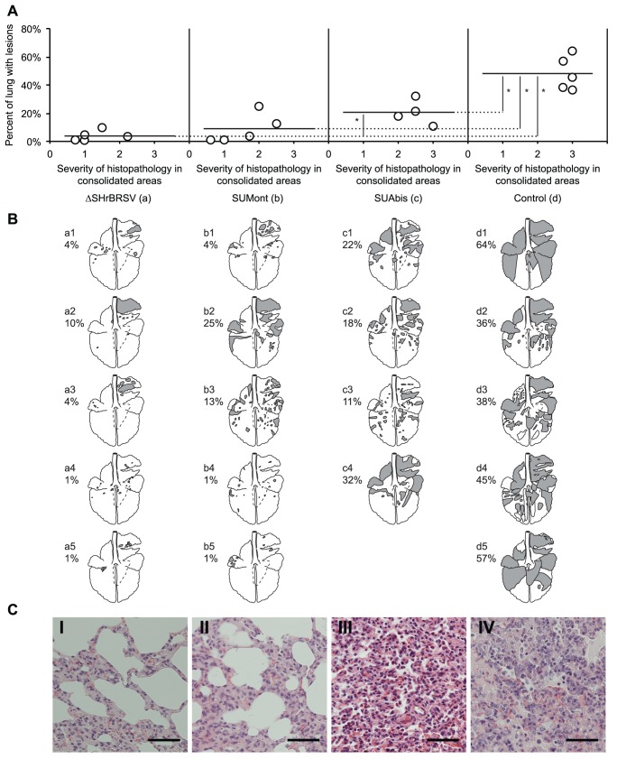
Figure 3. Vaccination reduces the extent of lung lesions following BRSV challenge.
Four groups of 5 calves were vaccinated as described in Fig. 1 and challenged with BRSV, 5 weeks after vaccination. Two weeks before challenge, one calf (c5) was euthanized due to traumatic injury. Lungs were removed after exsanguination, lesions were recorded on a lung chart after visual examination and palpation, and the proportion of lung showing pneumonic consolidation was calculated. Formalin-fixed tissue samples from each lobe in the right lung were analyzed for the severity of histopathological changes and scored as either normal (0), mild (1), moderate (2) or severe (3). (A) shows the extent of macroscopic lesions on the y-axes, and the microscopic severity of inflammation (mean score of four sections per calf) on the x-axes. Statistically significant difference is indicated by asterisks (p≤0.05). (B) shows the percent of pneumonic consolidation in each animal (also depicted as filled areas in lung-charts), and emphysema (outlined areas in calves d3 and d4). Panels C (I–IV) show representative histological images from each of the four groups of calves. Bar indicate 100 µm. Panels C (I) (ΔSHrBRSV), C (II) (SUMont), C (III) (SUAbis) and C (IV) (Control) show lung parenchyma with minimal, mild, moderate and severe pathological changes, respectively.

