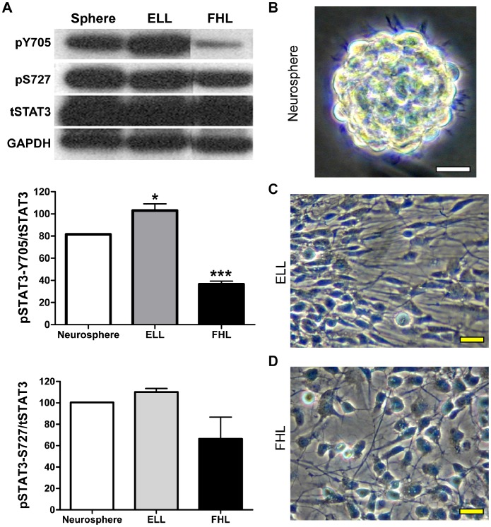
Figure 1. EGF and FGF2 have opposite effects on STAT3 phosphorylation in hNSCs.
(A) Lysates from neurospheres as well as hNSCs primed with either EGF (ELL) or FGF2 (FHL) for 24 hrs were analyzed by immunoblotting using antibodies against phosphorylated STAT3-Tyr705 (pY705) or STAT3-Ser727 (pS727). The blots were stripped and re-probed for total STAT3 (tSTAT3) and GAPDH (loading control). Densitometric analysis was performed and ratios of phosphorylated STAT3 to total STAT3 (pSTAT3/tSTAT3) are graphically represented as Mean ± SEM (n = 3). *p<0.05 and ***p<0.001 by One-way ANOVA with Tukey post-hoc test. pSTAT3-Y705 is increased in EGF-primed cells, but reduced in FGF2-primed cells when compared to unprimed neurospheres. pSTAT3-S727 levels, however, remain unchanged after either priming. Representative phase contrast images of primary culture hNSCs are shown as neurospheres (B), one day after ELL- (C) or FHL-priming (D). Scale bars, 20 µm. hNSCs: human neural stem cells; ELL: EGF plus LIF and laminin; FHL: FGF2 plus heparin and laminin.

