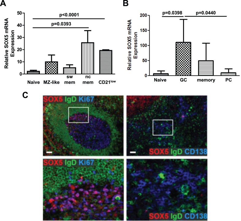
Figure 3. Expression of SOX5 in human B cell subpopulations.
(A) Relative quantification of SOX5 by RT-qPCR in peripheral blood naive, MZ-like, switched memory (sw mem), non-classical memory (nc mem) and CD21low B cells. (B) Relative quantification RT-qPCR assay for SOX5 expression in follicular naive, germinal center B cells (GC), memory B cells and plasma cells (PC) from tonsils. T-test p-values indicate the significance of differences between the samples. Relative expression levels of SOX5 are shown as mean ± SD. RPLP0 gene was used as an internal control in the samples. (C) Immunofluorescence staining for the expression of SOX5 protein in tonsillar tissues. IgD staining was used to stain mantle zones, Ki67 staining for proliferating centroblasts within germinal centers and CD138 as a marker for extrafollicular plasma cells.

