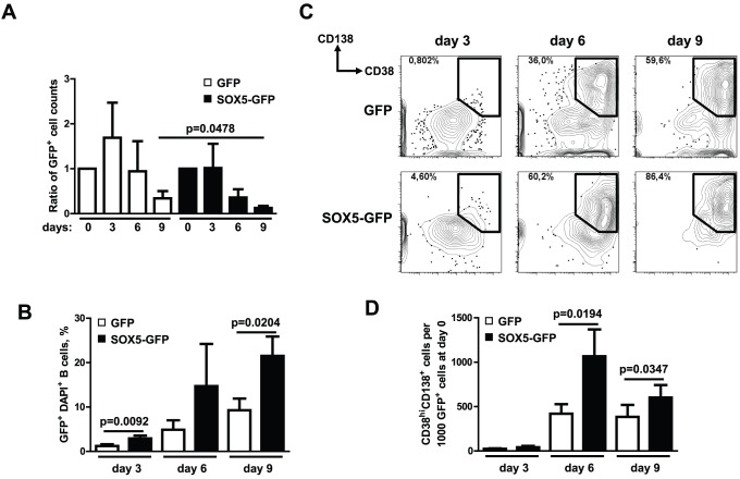
Figure 6. SOX5 modulates in vitro terminal B cell differentiation.
(A) Proliferation measured as the ratio of absolute numbers of GFP+ cells in the samples. Absolute cell counts of GFP+ cells on day 0 are taken as 1.0 and the data are expressed as mean ± SD. Summary of three independent experiments are depicted. (B) Frequencies of GFP+ DAPI+ cells within GFP (control) and SOX5-GFP-transduced peripheral blood B cells cultivated in vitro. (C) Plasma cell differentiation analyzed by FACS in peripheral blood B cells stably transduced either with GFP (control) or SOX5-GFP fusion construct upon in vitro stimulation with IL4+CD40L+IL21. DAPI-negative GFP+ gated cells were analyzed by FACS for the plasma cell markers CD138 and CD38 at days 3, 6 and 9. Gates indicate the frequencies of CD138+CD38hi plasmablasts in each FACS plot. Representative FACS plots of three independent experiments are shown. (D) Numbers of CD138+CD38hi plasmablasts in GFP (control) and SOX5-GFP-transduced B cells at days 3, 6 and 9 referring to CD38hi CD138+ cells per 1000 GFP+ cells at day 0. Cell numbers are depicted as mean ± SD and t-test p-values indicate the significant differences. Summary of three independent experiments are shown.

