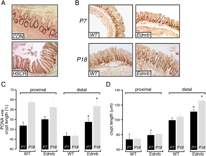
Figure 1. Colon crypt proliferation in Ednrb−/− mice.
A. Representative images showing PCNA staining of crypt cells from human distal colon from control (CON) and HSCR patients. B. Representative images showing PCNA staining of crypt cells from distal colon of P7 and P18 WT and Ednrb−/− mice. C. Summary graph showing extent of PCNA-immunoreactive zone (% of total crypt length that is PCNA-positive) in the epithelium of proximal and distal colon from WT (n = 5) and Ednrb−/− mice (n = 5). D. Mean crypt length in proximal and distal colon from WT (n = 5) and Ednrb−/− mice (n = 5). *p<0.05.

