Figure 2.
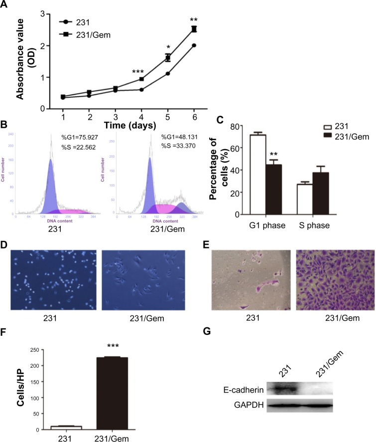
231/Gem cells display increased proliferation and invasion.
Notes: (A) Cell proliferation in 231 and 231/Gem was detected by CCK-8. *P<0.05, **P<0.01, ***P<0.001. (B) Cell cycle analysis was performed using flow cytometry by staining with propidium iodide. (C) Columns represent quantification of cell phase. **P<0.01. Inverted microscopy (D) and Transwell chambers (E) were used to evaluate the alteration of morphology and invasiveness of 231 and 231/Gem cells. (F) Quantification of invasion assays. Cells were counted in triplicate wells and in three identical experiments. ***P<0.001. Columns represent the means of three independent experiments, error bars represent SD. (G) Expression of cell adhesion-associated protein E-cadherin was assessed using Western blot.
Abbreviations: 231, human breast cancer cell line MDA-MB-231; 231/Gem, chemoresistant human breast cancer cell line MDA-MB-231/gemcitabine; CCK-8, Cell Counting Kit-8; GAPDH, glyceraldehyde 3-phosphate dehydrogenase; HP, high-power objective; OD, optical density; SD, standard deviation.

