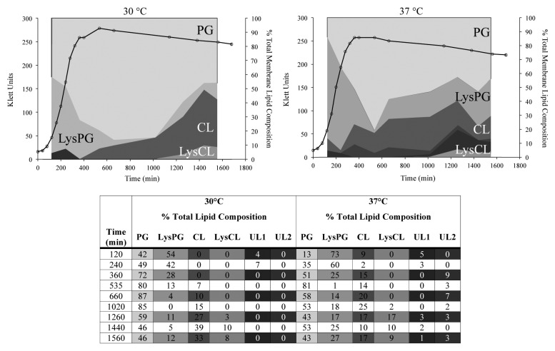
Figure 1. Temperature-dependent changes in the membrane lipid composition of L. monocytogenes. Growth of L. monocytogenes EGD-e in BHI in the presence of [32P] PPi is represented as a measure of optical density by the black line. Aliquots of bacterial culture were removed and used for lipid isolation and subsequent separation by TLC. The percentage of each lipid species of the total lipid composition was determined by phosphorimaging. The percent of the total lipids made up of each species is relative to the area occupied on the graph. Detected lipids include cardiolipin (CL), lysylcardiolipin (LysCL), lysylphosphatidylglycerol (LysPG), phosphatidylglycerol (PG), unidentified lipid 1 (UL1), and unidentified lipid 2 (UL2). UL2 was present in the membranes of L. monocytogenes EGD-e grown at 37 °C (B), but not at 30 °C (A).

An official website of the United States government
Here's how you know
Official websites use .gov
A
.gov website belongs to an official
government organization in the United States.
Secure .gov websites use HTTPS
A lock (
) or https:// means you've safely
connected to the .gov website. Share sensitive
information only on official, secure websites.
