
Figure 4.
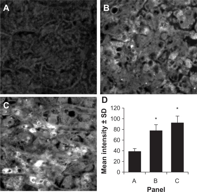
Immunofluorescence of HCC as detected by HCA519/TPX2.
Notes: Two archival HCC samples were re-cut and stained using the anti-HCA519/TPX2 antibody. (A) shows normal liver that did not stain with the anti-HCA519/TPX2 antibody. (B and C) illustrate positive staining for HCA519/TPX2. All magnifications were taken through the 40×. (D) presents the average fluorescent intensity of the three different panels from representative areas of the non-cancerous liver or HCC. For each panel, three distinctive representative areas were imaged, the intensity was quantitated, and the intensity is presented ± SD. *The difference (P<0.05) in fluorescence of the HCC compared to the non-cancerous tissue.
Abbreviations: HCC, human hepatocellular cancer; HCA519, hepatocellular carcinoma-associated antigen-519; SD, standard deviation; TPX2, targeting protein for Xklp-2.