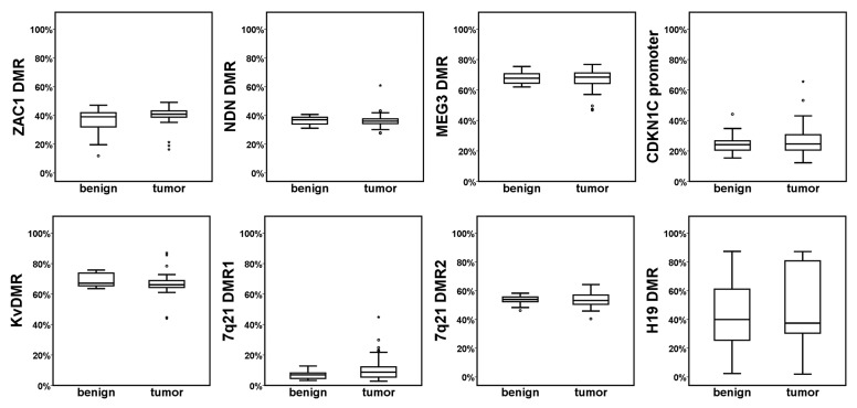
Figure 3. Methylation of imprinted DMRs in benign and tumor prostatic tissues. DNA methylation (%) of the indicated imprinted DMRs in benign and tumor prostatic tissues was quantitated by bisulfite pyrosequencing. The boxplots represent the mean methylation values of all assessed CpG positions for the indicated region for benign or tumor samples. Mann-Whitney-U test was used to evaluate the differences between the two sample groups; none of the differences was significant at P < 0.05. Note the exceptionally large variation in the H19 DMR.

An official website of the United States government
Here's how you know
Official websites use .gov
A
.gov website belongs to an official
government organization in the United States.
Secure .gov websites use HTTPS
A lock (
) or https:// means you've safely
connected to the .gov website. Share sensitive
information only on official, secure websites.
