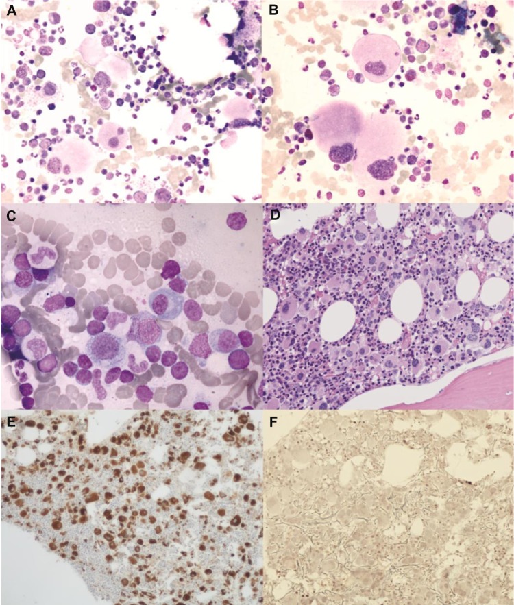
Figure 1.
(A and B) Light microscopy examination of marrow aspirate at baseline (May Grünwald–Giemsa) showing atypical, hypolobated megakaryocytes (A: original magnification ×50; B: original magnification ×100). (C) After 6 months of lenalidomide therapy, abnormal megakaryocytes disappeared and a scattered lymphocyte infiltration was seen (original magnification ×100). (D–F) Bone marrow trephine image at baseline demonstrating increased cellularity with numerous atypical hypolobated megakaryocytes (D: original magnification ×20, hematoxylin–eosin; E: original magnification ×10, immunoperoxidase staining for von Willebrand factor). Only a few reticulin fibers were present (F: original magnification ×20, reticulin staining).

