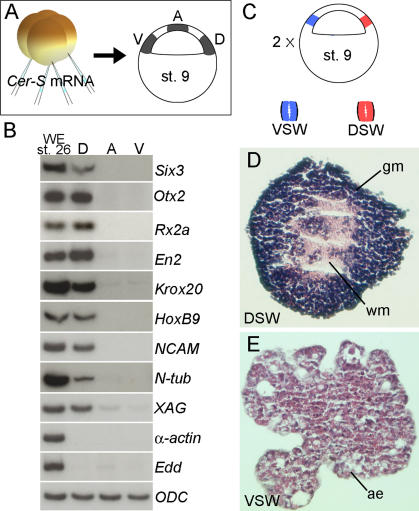
Figure 3. The Blastula Dorsal Animal Cap Is Specified to Form CNS.
(A) Experimental diagram showing embryos injected with CerS mRNA from which three regions of the animal cap were dissected at blastula, cultured until stage 26, and processed for RT-PCR. The size of the explants was 0.3 mm by 0.3 mm in these samples. Abbreviations: A, animal pole; D, dorsal region; V, ventral animal cap.
(B) RT-PCR analysis of animal cap fragments; note that anterior brain markers were expressed in the dorsal fragments in the absence of mesoderm (α-actin) and endoderm (endodermin, Edd) differentiation. Abbreviations: A, animal pole; D, dorsal region; V, ventral animal cap.
(C) Experimental diagram of the small animal cap sandwich experiments; these embryos were not injected with CerS. In this case, the size of the explants was 0.15 mm by 0.15 mm leaving a 0.15-mm gap from the floor of the blastocoel to avoid contamination from mesoderm-forming cells. Fragments from two explants were sandwiched together (explants are too small to heal by curling up) and cultured in 1× Steinberg's solution until stage 40. Abbreviations: VSW, ventral sandwich; DSW, dorsal sandwich.
(D) Histological section of dorsal animal cap explant (dorsal sandwich). These sandwiches differentiated into histotypic forebrain tissue including white and gray matter (4/17). Abbrevations: DSW, dorsal sandwich; gm, gray matter; wm, white matter.
(E) Histological section of a ventral animal cap sandwich. All sandwiches differentiated into atypical epidermis (n = 20). Abbreviations: ae, atypical epidermis; VSW, ventral sandwich.

