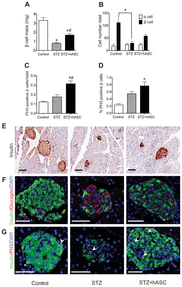
Figure 2. Systemic administration of hASCs improves islet morphology and β cell mass in STZ-treated NOD-SCID mice.
A: Average β cell mass in PBS-treated controls, STZ-treated, and STZ+hASC-treated mice. B: Average cell number of glucagon positive cell (α cells) and insulin positive cells (β cells) per islet at day 17. C–D: PH3 was used to detect proliferating β cells. C: Number of PH3 positive β cells per islet. D: Percentage of PH3 positive β cells. Results are expressed as the means ± S.E.M; n=4 mice per group. *p<0.05 compared to Control; #p<0.05 compared to STZ-treated mice. E–G: Representative images of pancreata from indicated treatment group 7 days after hASC administration. Scale bars = 100 μm. E: Insulin immunocytochemistry shown at 20X magnification. F: Immunofluorescent detection of insulin (green) and glucagon (red) in islets of treated mice (40X magnification). G: Immunofluorescent detection of insulin (green) and PH3 (red) shown at 40X magnification. Arrows depict cells double positive for insulin and PH3.

