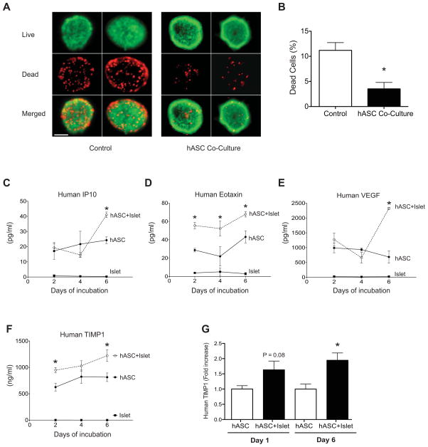
Figure 3. Co-culture with human ASCs improves islet viability, while human ASCs co-cultured with islets secrete TIMP-1 in excess of other paracrine factors.
A: Live dead staining of C57BL6/J mouse islets co-cultured with (hASC Co-Culture) or without (Control) hASCs in a transwell system for six days. Representative fluorescent images of dead, ethidium homodimer-1 positive cells (red) and viable, calcein-AM positive cells (green) are shown at 20X magnification (scale bar = 50 μM). B: Quantification of the percentage of dead cells in dispersed islets cultured alone or with hASCs. *p<0.05 compared to islets cultured without hASCs. Results are expressed as the means ± S.E.M, n=3 independent experiments. C–F: ELISA and multiplex analysis of factors secreted by hASCs after 2, 4, and 6 days of islet co-culture. Supernatant concentration of human IP10 (C), human eotaxin (D), human VEGF (E), and human TIMP-1 (F). hASC alone = ●, hASC+C57BL6/J islets = ○, mouse islets alone = ■. Note, concentration of assayed factors is expressed as pg/mL except for TIMP-1, which is expressed as ng/ml. G: hASCs and human islets were co-cultured for 6 days in a transwell system. Supernatant concentration of human TIMP-1 was measured at day 1 and 6. *p<0.05 compared to hASCs cultured alone. Results are expressed as the means ± S.E.M. n=4 per group.

