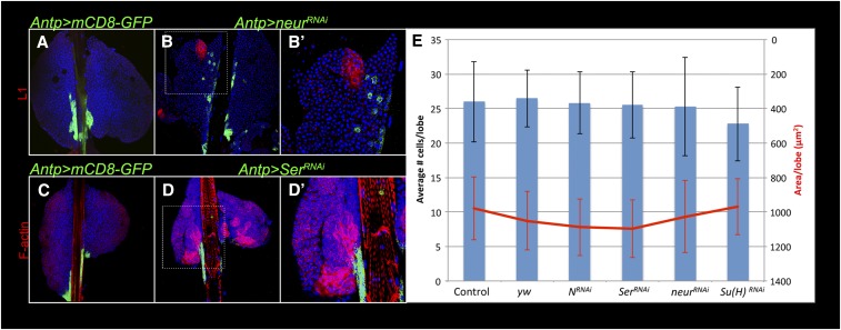
Figure 9.
Lamellocyte differentiation is induced by neurRNAi and SerRNAi. (A) Control Antp > GFP and (B and B′) Antp > neurRNAi lymph glands stained with anti-L1 antibody (red). L1-positive lamellocytes are found in the cortex adjacent to satellite niche cells (green). (C) Control Antp > GFP and Antp > SerRNAi (D and D′) lymph glands stained with rhodamine-phalloidin. F-actin-rich lamellocytes (red) are adjacent to the GFP-positive niche cells. Hoechst stains DNA (blue). (E) Niche size is unaffected by knockdown of N pathway components. Left: Average number of GFP-positive cells per niche (n ≥ 20) are not significantly different in control (Antp > mCD8-GFP and Antp > y w) lobes and experimental (RNAi of N, Ser, neur or Su(H)) lobes. Right: The average area of GFP-positive cells per niche (n ≥ 20) is not significantly different between control and experimental lymph glands. Standard deviations for both parameters are shown.

