
Figure 2.
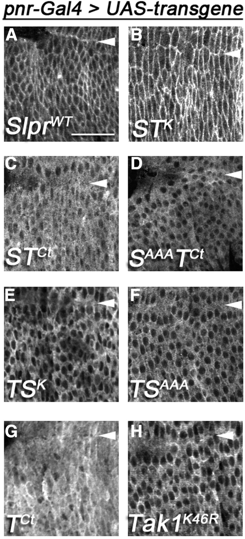
Differential localization of transgenic proteins in embryonic dorsal epidermis maps to the C terminus. (A–G) Anti-HA and (H) anti-Tak1 immunostaining. The indicated constructs were expressed in the embryo with the pnr-Gal4 driver. Images are single confocal slices ∼2 μm below the apical surface of the epidermis. Views are dorsolateral, surrounding the posterior canthus of the zippering epidermis during dorsal closure in stage 15 embryos. Arrowheads indicate the dorsal midline. Bar, 20 μm.