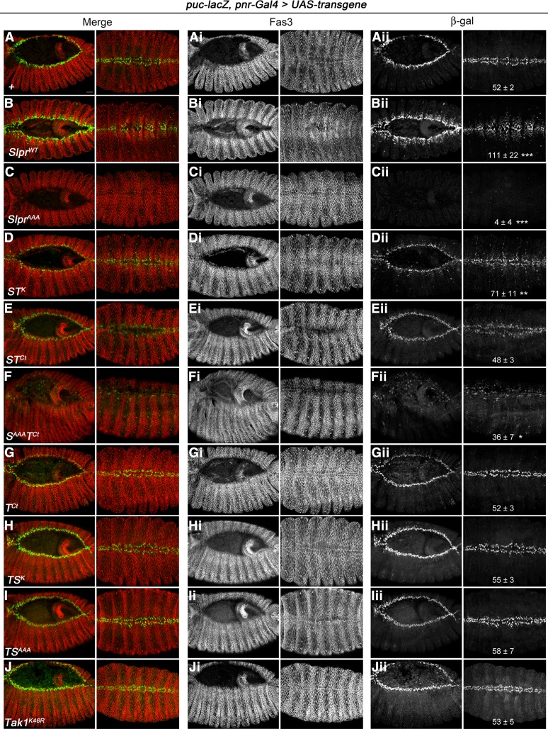
Figure 5.
Specificity of Slpr vs. Tak1 signaling in activation of JNK target gene expression during dorsal closure. Early and late progression of dorsal closure (stage 13–14, left; stage 15, right) is shown in merged panels (A–J) and in individual channels, with immunostaining for either Fas3 (Ai–Ji) or β-gal to detect puc-lacZ enhancer trap expression (Aii–Jii). Transgenes indicated in the lower left of each panel (A–J) are expressed in the dorsal ectoderm and amnioserosa under the control of pnr-Gal4. Embryos are shown dorsally with anterior to the left. Bar, 20 μm. Quantification of puc-lacZ in stage 15 embryos as a proxy for JNK pathway activity is given in the rightmost panels as the mean number of β-gal positive nuclei per five hemisegments ± SD based on 4–8 embryos. Significant differences compared to the no Tg control (Aii) are indicated based on one-way ANOVA using Bonferroni’s multiple comparisons test vs. the control. ***P < 0.005, **P < 0.01, *P < 0.05.

