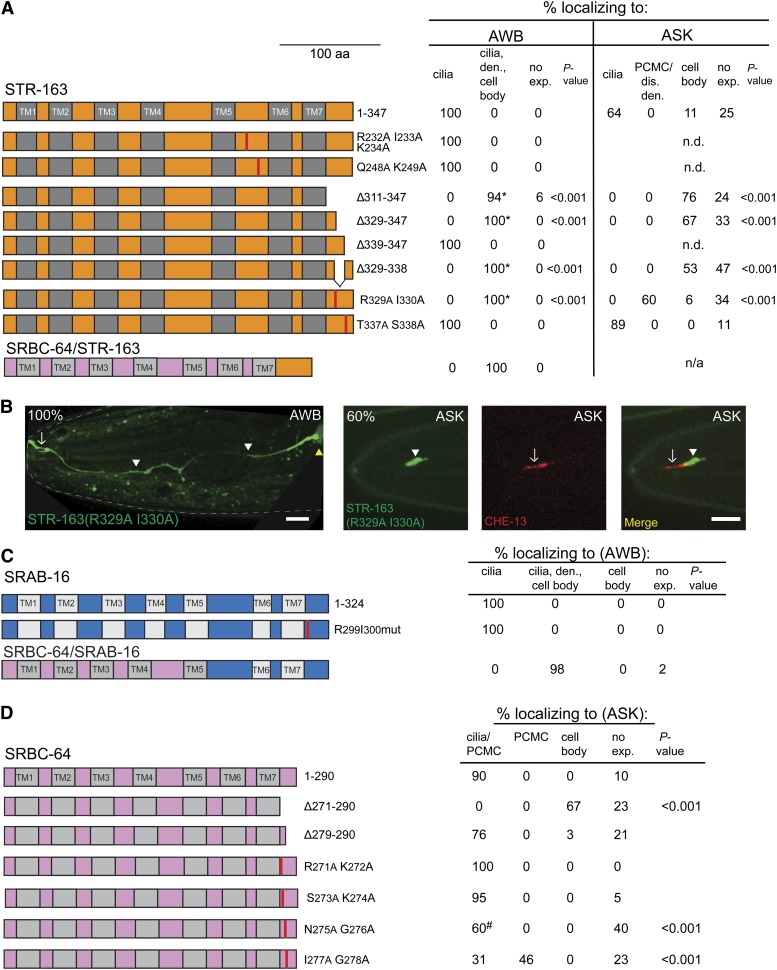
Figure 3.
Identification of novel sequences required for ciliary localization of GPCRs in AWB or ASK. (A) Localization patterns of full-length and indicated mutant STR-163::GFP fusion proteins in AWB and ASK. The localization pattern of a SRBC-64/STR-163::GFP chimeric fusion protein in AWB is also shown. GFP coding sequences were fused to the C termini of the indicated proteins. Sequences mutated in the third intracellular loop are shown in Figure S2. *, localization to the membranes of the indicated cellular compartments; vertical red lines, position of mutated residues; TM, transmembrane domain; den., dendrite; dis. den., (includes ciliary base) dendrite; PCMC, periciliary membrane compartment; no exp., no expression; n.d., not done; n/a, not applicable. n = 30–60 animals each; two independent transgenic lines were examined for each construct. (B) Localization of mutant STR-163::GFP fusion proteins in AWB (left panel) and ASK (right panels). ASK cilia were visualized via cell-specific expression of CHE-13::TagRFP. White arrows indicate cilia; white arrowheads indicate dendritic domains/PCMC; yellow arrowheads indicate cell bodies. Bars, 10 μm (left panel), 5 μm (right panels). (C) Localization pattern of full-length, chimeric, and mutant SRAB-16::GFP in AWB. Vertical red lines indicate position of mutated residues. n = 20–35 animals each; two independent transgenic lines were examined for each construct. (D) Localization of full-length and mutant SRBC-64::GFP fusion proteins in ASK. Vertical red lines indicate position of mutated residues. #, weak expression. n = 20–35 animals each; two independent transgenic lines were examined for each construct.

