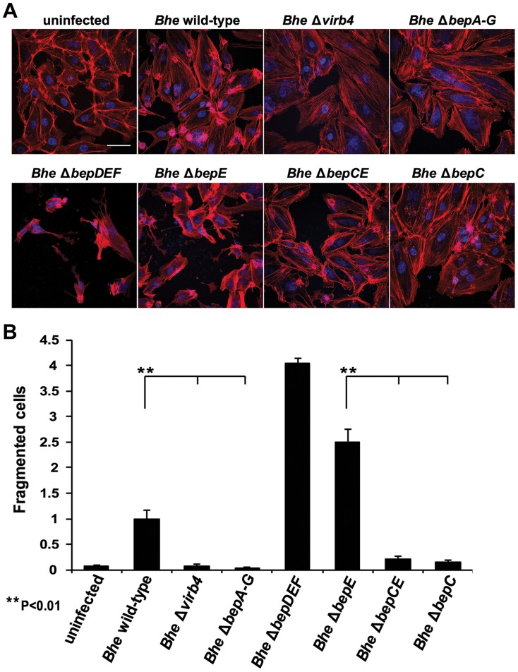
Figure 4. The double deletion mutant Bhe ΔbepCE abolishes cell fragmentation.
(A) Subconfluent monolayers of HUVECs were infected for 48 h with MOI = 200 of the Bhe strains depicted in the figure or were left uninfected. Samples were then fixed, stained immunocytochemically and analyzed by confocal laser scanning microscopy. F-actin is represented in red (Phalloidin) and DNA in blue (DAPI) (scale bar = 50 µm). (B) Quantification of cell fragmentation at 48 h post infection was performed as described for Fig. 1C and D and presented as mean of triplicate samples +/− SD. Statistical significance was determined using Student's t-test. P<0.05 was considered statistically significant. Data from one representative experiment (n = 3) are presented.

