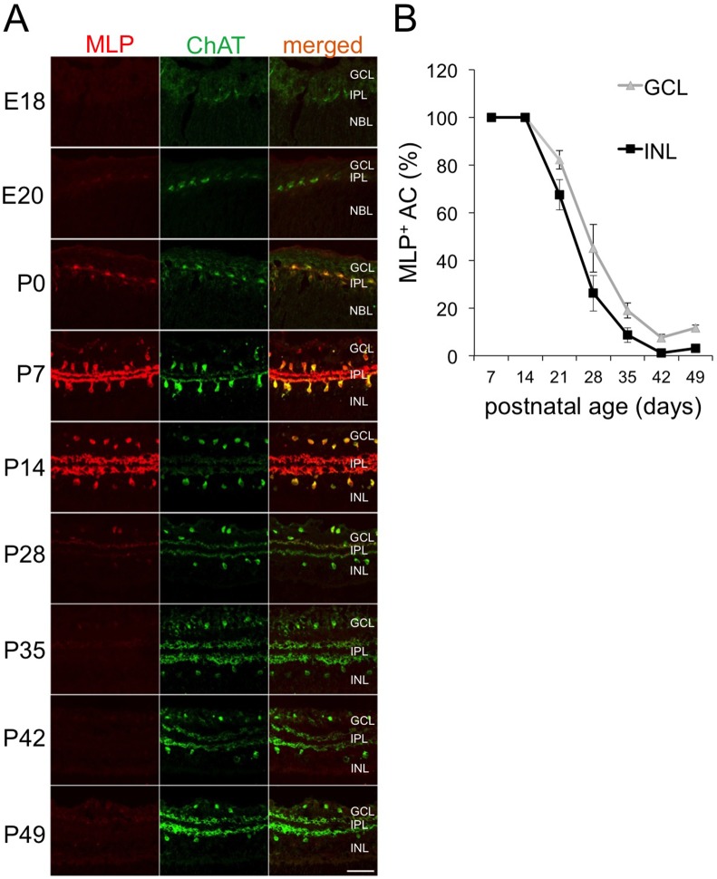
Figure 2. Expression of MLP in postnatal ChAT-positive AC.
A: Confocal images were taken of E18, E20, P0, P7, P14, P28, P35, P42 and P49 retinae after co-staining with antibodies against ChAT and MLP. MLP expression becomes detectable at E20 in ChAT-positive AC located in the inner nuclear layer as well as ganglion cell layer. Levels reached a peak between P7 and P14. MLP expression dramatically decreases afterwards. GCL = ganglion cell layer, IPL = inner plexiform layer, INL = inner nuclear layer Scale bar: 50µm. B: Quantification of MLP-positive cholinergic AC in the inner nuclear layer (INL) and ganglion cell layer (GCL, displaced) of retinae of 7–49 days old rats. All cholinergic AC were positive for MLP at P7 and P14. Numbers of MLP-positive AC continuously decreased between P21 and P42. This attenuation proceeded slightly faster in the INL than in the GCL.

