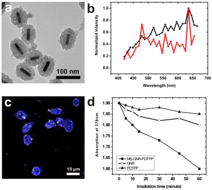
Figure 3.
(a) TEM imaging of MS-GNR-PdTPPs; scale bar: 0.10 µm (b) Energy transfer in MS-GNR-PdTPPs, showing the TPLs of MS-GNRs (black) and MS-GNR-PdTPPs (red). (c) Confocal microscopy imaging of the endocytosis of MS-GNR-PdTPPs, with the nuclei stained by Hochest 33342 (blue signal); white foci are the TPLs of MS-GNR-PdTPPs under 800 nm laser excitation; scale bar: 10 µm. (d) Decay of optical absorption of ADPA at 378 nm, caused by the generation of singlet oxygen from GNRs (○), PDTPPs (▲) and MS-GNR-PdTPPs (■) as a function of laser irradiation exposure time.

