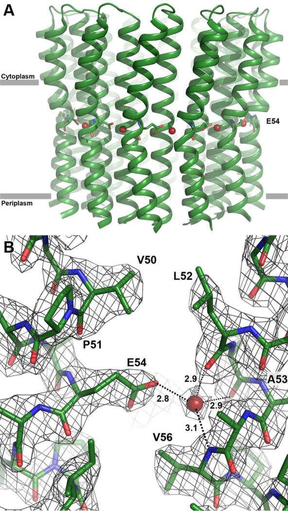
Figure 1. Structure at 2.4 Å resolution of the B. pseudofirmus OF4 c13 ring at pH 9.0.
The view is from the membrane plane. E54 is shown as sticks; the red spheres indicate the water molecules in the ion-binding sites. (A) Overview of the c13 ring with cytoplasmic side on top in ribbon representation. (B) Close-up of the ion-binding site. E54 is protonated and in locked conformation, despite the high pH. The 2Fobs-Fcalc electron density map is shown as a grey mesh at 1.8σ.

