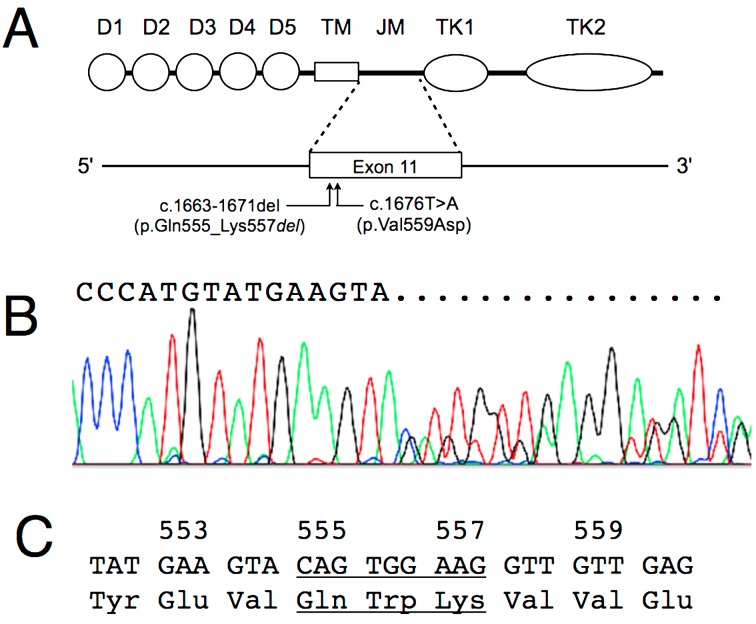
Fig. 2.
A: Diagram of KIT domain structure and exon 11 indicating position of mutations in Case 1 and Case 2. Both are located in proximal region of exon 11. Detail mutation of p.Val559Asp is shown in Fig. 3. D1 to D5, immunoglobulin-like domains; TM, transmembrane domain; JM, juxtamembrane region; TK1, adenosine triphosphate binding domain; TK2, kinase activation loop. B: Sequence analysis of the exon 11 amplicon from Case 1. An overlapping curve is seen in the sequencing result, showing a 9-bp deletion mutation. C: The nucleotide and amino acid sequences of c-KIT exon 11 from Case 1. The 9-bp deletion mutation, located 19 bases downstream of the first codon of exon 11, and the deleted amino acids are underlined.

