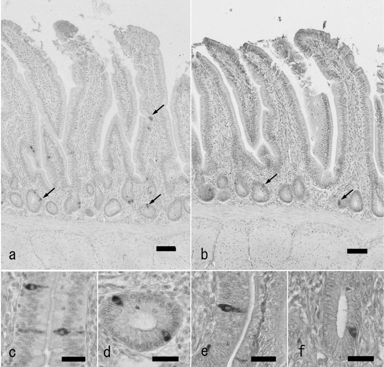
Fig. 1.
GLP-2-immunoreactive cells in the chicken distal ileum of antigen retrieval agent-treated (a, c, d) and untreated sections (b, e, f). a: Localization of GLP-2-immunoreactive cells (arrows) in the distal ileum of antigen retrieval agent-treated section. Immunoreactive cells are located in the epithelium of the lower part of villi and crypts mainly. Bar: 50 µm. b: Localization of GLP-2-immunoreactive cells (arrows) in the chicken ileum of untreated sections. A small number of immunoreactive cells (arrows) are observed. c, d: High magnification views of GLP-2-immunoreactive cells in villous epithelium (c) and crypt (d) of antigen retrieval agent-treated sections. Bar: 20 µm. e, f: High magnification views of GLP-2-immunoreactive cells in villous epithelium (e) and crypt (f) of untreated sections. Bar: 20 µm.

