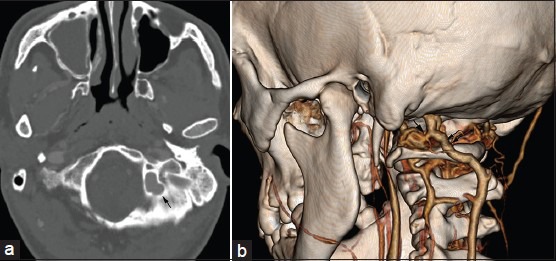
Figure 2.

Multiplanar image reformatting (a) and 3D volume rendered (b) Images of the same patient show that the left large posterior condylar vein courses along the posterior condylar vein canal (arrow in a), originates from the superior bulb of the internal jugular vein, and drains into the deep cervical vein (double arrows in b). It connects with the horizontal portion of the vertebral artery venous plexus (arrow in b)
