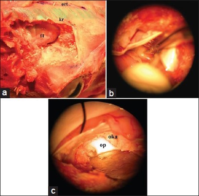
Figure 3.

(a) Following the craniectomy to simulate the standard frontal craniotomy and dura was opened in a semicircular fashion. This way it is possible to simulate the standard frontal approach in the human brain (b) The next step is identification and microsurgical dissection of the optic nerve. The filled arteries in Sylvian dissection step allows to feel a real surgery experience for inexperienced neurosurgery residents (c) At the end of the dissection, the last step is the opening of optic canal and exposure of the optic nerve within the canal
