Fig. 9.
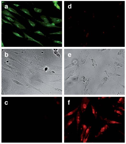
Ru-catalyzed deprotection of 10 μM ©5 in CEF cells. Left column: ©5, 30 min at 37 °C; (a) green channel; (b) brightfield; (c) red channel. Right column: ©5 incubated with 20 μM [Ru] and 100 μM PhSH, 20 min; (d) green channel; (e) brightfield; (f) red channel. Fluorescence microscopy settings for green channel: filter 530–550 nm, emission filter 590 nm and dichromatic mirror 570 nm; and for red channel: filter 530–550 nm, emission filter 590 nm and dichromatic mirror 570 nm.

