
Fig. 2.
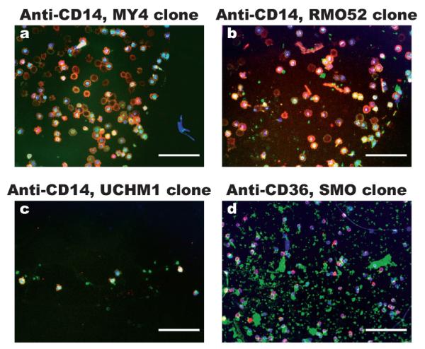
Representative images of cells captured from erythrocyte lysed blood onto antibody spots under static incubation conditions. The antibodies used are (a) anti-CD14 clone MY4, (b) anti-CD14 clone RMO52, (c) anti-CD14 clone UCHM1 and (d) anti-CD36 clone SMO. After rinsing off unbound cells, the cells captured on the antibody spots were stained using FITC-anti-CD36 (green), PE-anti-CD14 (red) and DAPI (blue). The images were created by overlapping the fluorescence photographs. The triple stained cells are monocytes, CD36 + CD14− cells are likely platelets and CD36−CD14+ cells are likely neutrophils. From the images, anti-CD14 MY4 clone and RMO52 clone captures monocytes efficiently, while anti-CD36 captures mostly platelets. (Scale bars: 100 μm).