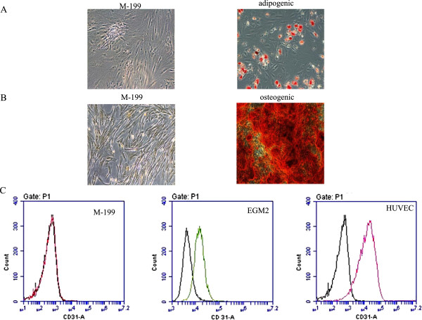
Figure 1.

ASC differentiation to adipose, osteocyte and endothelial like cells. Oil Red O staining was used to detect lipid droplet formation during adipogenic differentiation (panel A). The left side image depicts control ASCs grown in M-199 medium while the right one shows ASCs grown in adipogenic differentiation medium. Alizarin Red staining was used to measure calcium deposition during osteogenic differentiation (panel B). The left side image depicts control ASCs grown in base medium M-199 while the right one shows ASCs grown in osteogenic differentiation medium. Endothelial differentiation was measured by CD31 flow cytometry (panel C). Left side is undifferentiated ASCs grown in M-199 medium, the middle is differentiated ASCs grown in EGM2 medium and the right is HUVECs endothelial cell control.
