
Figure 1. Virtual docking of MDP and SE ligands on CRT.
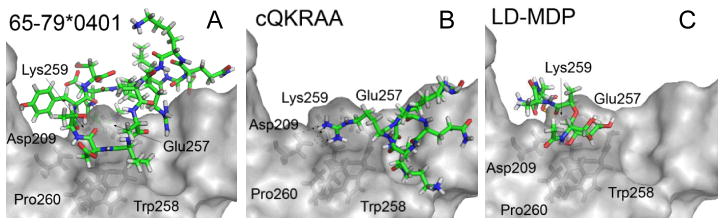
Ligands are shown as colored sticks; CRT surface is in light gray; CRT residues in close proximity to ligands are shown as dark-gray sticks. As can be seen, the two SE ligands, 15-mer linear peptide 65–79*0401 (A) and the SE-mimetic cQKRAA (B), as well as LD-MDP (C), are predicted to occupy overlapping binding sites on CRT, in significant proximity to several key CRT residues.