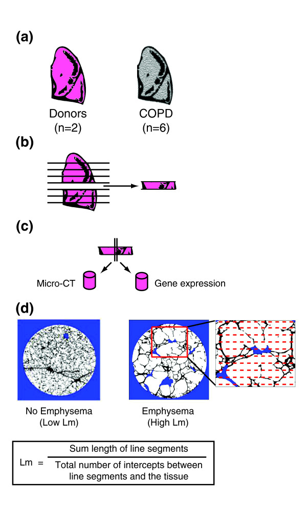
Figure 1.

Outline of study design. (a) Whole lungs were removed from patients with severe COPD and from donors, inflated with air, and rapidly frozen in liquid nitrogen vapor. (b) The frozen specimens were cut into 2-cm slices from apex to base of the lung. (c) Adjacent tissue cores were removed from 8 different slices of each lung (8 patients with 8 slices = 64 total regions). (d) Micro-CT was used to measure Lm at 20 evenly spaced intervals throughout one core from each region.
