
Figure 3.
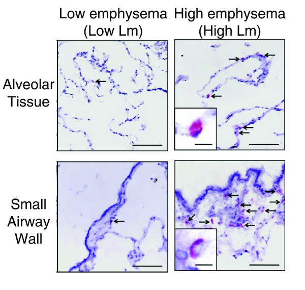
Validation of differential expression for CD79A by immunohistochemistry. Representative images of CD79A-positive cells (arrows) in the alveolar tissue and the small airway walls. Positive staining appears red. Scale = 200 µm; inset = 10 µm.