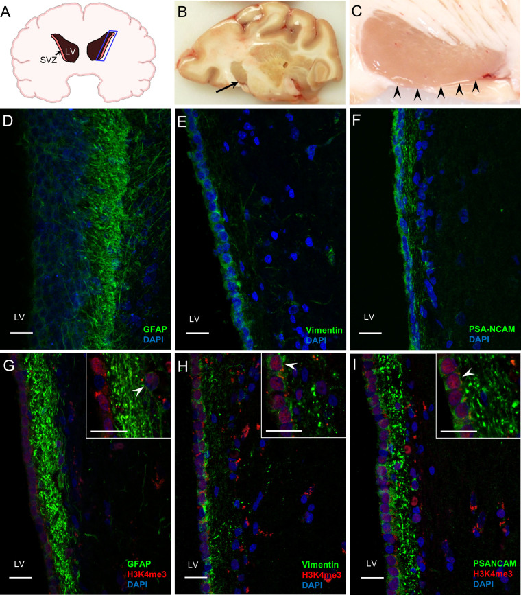
Figure 1. Gross anatomy of baboon SVZ architecture and the colocalization of H3K4me3 and cell type specific markers in baboon SVZ.
(A) Scheme displays coronal view of baboon brain with dictation of lateral ventricle (LV) and subventricular zone (SVZ). (B) Coronal slice of baboon forebrain showing regions sampled, the arrow points to the head of the caudate nucleus at the margin with the lateral ventricle. The dorsal is to the left and medial is at the bottom for orientation. 2× magnification. (C) Horizontal slice through the caudate nucleus showing the subventricular zone (SVZ) in temporal horn of lateral ventricle; arrow heads point to SVZ; 6× magnification. (D) Images of the caudal ventral subventricular zone reveal the GFAP-positive astrocytic ribbon. A deeper z-stack imaging was performed in order to view GFAP-positive processes of the NSCs extending toward the lateral ventricle (LV). (E) Vimentin staining shows NSC population in the baboon SVZ. (F) PSA-NCAM staining shows neuroblast population in the baboon SVZ. (G) A small population of H3K4me3 positive cells is colocalized with GFAP in rostral ventral SVZ. (H) Significant population of Vimentin-positive cells is colocalized with H3K4me3. (I) H3K4me3 persists in the PSA-NCAM positive neuroblast population. (D–I) 40× images; inserts are 100× magnification; White arrow heads in G and H indicate colocalization of H3K4me3 and cell type markers; (G–I) Images and inserts are 2um single slices. Scale bars present 20 μm.

