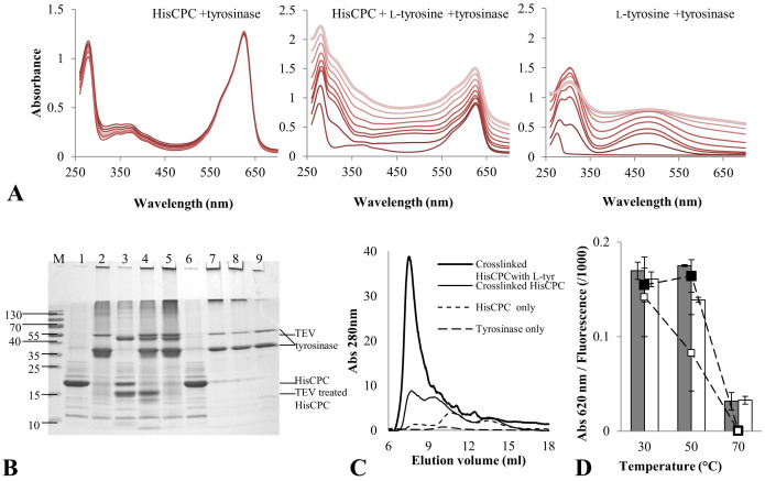
Figure 2. Tyrosinase catalyzed crosslinking of HisCPC.
(A) UV-Vis spectra monitoring the direct crosslinking of HisCPC (left panel) and crosslinking in the presence of L-tyrosine (middle panel) with tyrosinase VsTYR. For comparison, the oxidation of L-tyrosine with tyrosinase is also reported (right panel). Reactions were carried out in 100 mM K-phosphate pH 6.8 buffer. The periodic measurement of the absorbance spectrum is shown by shading from dark (initial conditions) to light grey (final conditions after 70 min). (B) SDS PAGE of HisCPC upon crosslinking in the presence or absence of L-tyrosine with tyrosinase. HisCPC (lane 1, 5 μg) was incubated in the presence of tyrosinase (lane 2), subjected to partial digestion with TEV (lane 3) and subsequently incubated in the presence of tyrosinase (lane 4), crosslinked with tyrosinase and subsequently incubated with TEV (lane 5), incubated with L-tyrosine (lane 6), and of both L-tyrosine and tyrosinase (lane 7). The product of incubation of L-tyrosine and tyrosinase (lane 8) and tyrosinase only (lane 9) are also shown). Molecular weight markers (lane M) are in kDa. (C) Size-exclusion chromatography of HisCPC in native and crosslinked form in the presence or absence of L-tyrosine. Separation was performed using a 17 ml volume column packed with Superdex 75 resin and a 1 ml/min flow of 100 mM potassium phosphate pH 7.5, 150 mM NaCl at 22°C. (D) Thermal stability of directly crosslinked HisCPC. Absorbance values at 620 nm (bars) and fluorescence values (square markers) were measured after 15 min incubation at different temperatures for native (filled bar and marker) and crosslinked (empty bar and marker) HisCPC.

