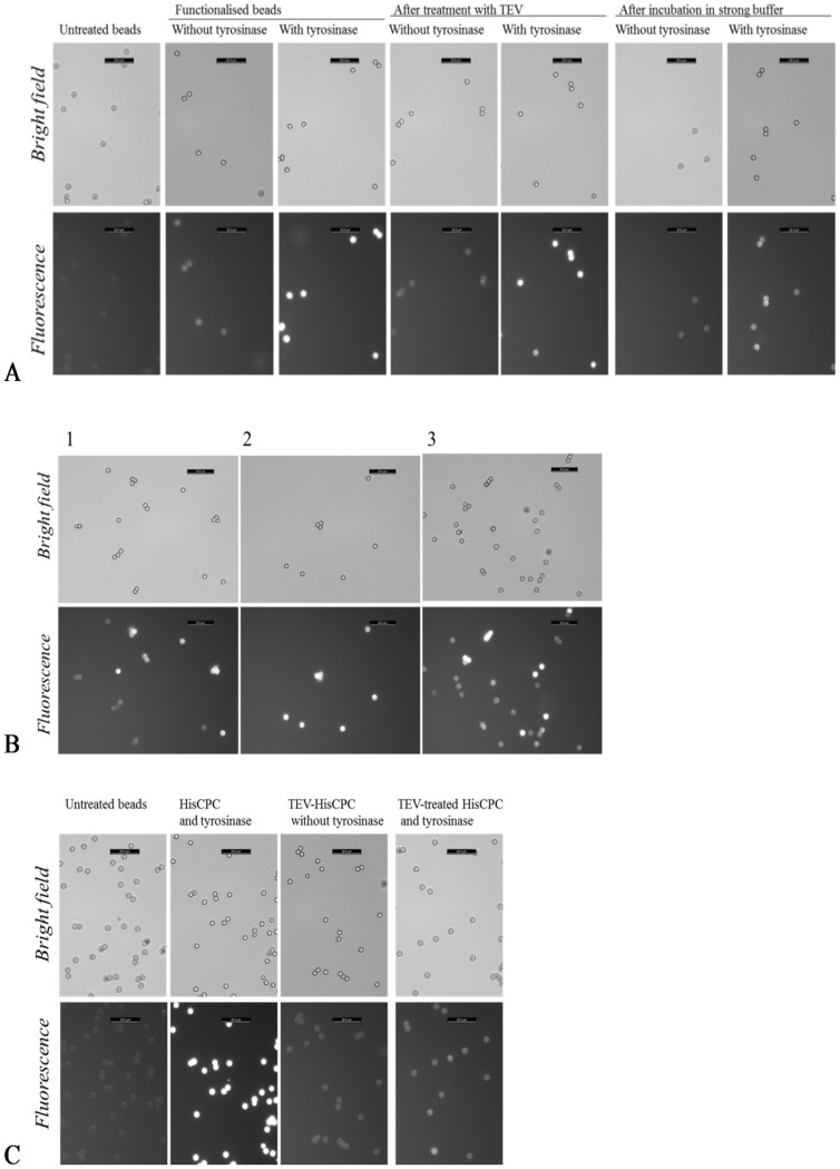
Figure 3. HisCPC immobilized on amino-modified beads using tyrosinase.
(A) Beads were first incubated in the presence of a partially purified HisCPC preparation and with and without tyrosinase, and then treated with either TEV-protease or a strong buffer containing 100 mM NaCl and 0.1% v/v Tween 20 for 4 h. (B) Microscopy images of combinations of differently treated amino-modified polystyrene beads. Sample 1, mix of adsorbed beads plus washed crosslinked beads. Sample 2, mix of crosslinked beads with and without washing. Sample 3, mix of adsorbed beads and cross-linked, protease treated beads. (C) Beads were incubated with tyrosinase and HisCPC full-length or previously treated with TEV-protease. The scale bar on the top right corner of each figure corresponds to 20 μm. Images were taken with a fluorescence microscope under bright light (top rows) and with fluorescence using a N21 filter (λex = 515–560 nm, λem ≥ 590 nm, bottom rows).

