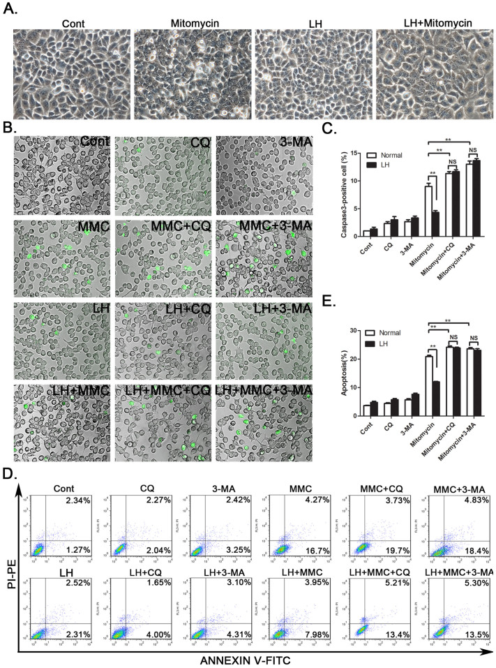
Figure 3. Chemotherapeutic agent-induced cell apoptosis was attenuated by autophagy in HCC cells.
(A). SMMC-7721 cells were treated with mitomycin (2 μM) under LH conditions for 24 hours. The morphology of the cells was recorded with a light microscope. (B). The fluorescent microscopic images of the apoptotic cells were captured via caspase3 staining. The quantification of the capase3-positive cells was described (C). (D and E). Annexin V staining and FACS analysis were performed after the mitomycin treatment for 24 h under normal or LH conditions in SMMC-7721 cells; the percentage of annexin V+ cells represents the apoptotic cells. MMC: Mitomycin. The experiments were repeated at least three times. **p < 0.01; NS, no significance.

